

Routine Chest Scans Hold Hidden Clues to Pulmonary Hypertension Survival, AI Study Reveals
Deep learning models extracted mortality predictors from standard CT imaging that remained significant even after accounting for established clinical risk scores, opening the door to retrospective risk stratification without additional radiation exposure.
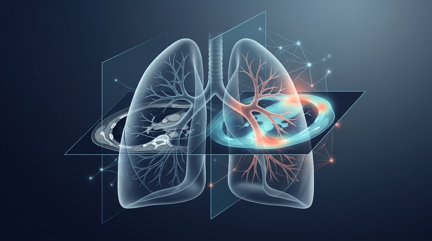
Artificial intelligence can extract powerful mortality predictors from routine chest CT scans in pulmonary hypertension patients—scans that are already sitting in hospital archives—according to research from the Sheffield Pulmonary Vascular Disease Unit in England. The findings suggest that standard imaging performed for diagnostic purposes could yield additional prognostic value without requiring specialized cardiac scans or exposing patients to additional radiation.
In a retrospective study of 620 patients from the ASPIRE registry enrolled between 2006 and 2020, researchers applied two validated deep learning models to non-gated, non-contrast chest CT scans. The AI systems measured cardiac chamber volumes and quantified coronary artery calcification—biomarkers that independently predicted death even after adjusting for the REVEAL 2.0 risk score, a comprehensive clinical tool already used to stratify pulmonary hypertension patients.
AI Uncovers Prognostic Signals in Standard Imaging
The research team deployed a cardiothoracic segmentation model built on the nnU-Net framework, trained on 50 non-contrast CT scans and validated on an external cohort of 55 scans. When tested on 368 scans, the model achieved area-under-curve values of 0.84 to 0.88, with sensitivity ranging from 70% to 94% and specificity from 63% to 100% across different cardiac structures.
A second model focused on coronary calcification detection demonstrated 95% sensitivity and 98% specificity compared to radiologist reports when validated on 473 scans. The AI identified calcification in 45% of right coronary arteries, 67% of mainstem or left anterior descending arteries, and 35% of left circumflex arteries among the study population.
The cohort included 472 patients with confirmed pulmonary hypertension and 148 without the condition. During follow-up, 388 deaths occurred (63% of participants), with mean survival of 1,659 days. Patients ranged across pulmonary hypertension subtypes: 202 with pulmonary arterial hypertension, 84 with left heart disease, 112 with lung disease-associated forms, 65 with chronic thromboembolic disease, and 9 with unclear classification.
Right Heart Size and Calcification Predict Mortality
In multivariable analysis adjusted for age, sex, and pulmonary hypertension subtype, right atrial volume emerged as a significant predictor, with each 10 mL increase associated with a hazard ratio of 1.024 (95% CI 1.009–1.039, p=0.001). Patients with right atrial volume exceeding 117.95 mL faced 61% higher mortality risk.
Coronary artery calcification showed even stronger associations. Mainstem or left anterior descending artery calcification carried a hazard ratio of 1.868 (95% CI 1.441–2.421, p<0.001), while right coronary artery calcification showed HR 1.378 (95% CI 1.113–1.705, p=0.003) and left circumflex calcification HR 1.342 (95% CI 1.084–1.660, p=0.007).
Critically, these AI-derived measurements retained prognostic significance even after adjusting for REVEAL 2.0 scores. Right atrial volume maintained a hazard ratio of 1.026 per 10 mL (95% CI 1.012–1.041), representing 2.6% higher risk per 10 mL increment. Coronary calcification remained strongly predictive: HR 1.604 for right coronary artery, 2.339 for mainstem/left anterior descending, and 1.576 for left circumflex (all p<0.001).
Among the 202 patients with pulmonary arterial hypertension specifically, the patterns held after REVEAL 2.0 adjustment, with left atrial volume showing HR 1.046 (95% CI 1.008–1.086, p=0.016) and right atrial volume HR 1.039 (95% CI 1.018–1.061, p<0.001).
Limitations and Future Directions
The study population was predominantly white British (80%), with mean age 64 years and 62% female representation. Most scans (78%) came from GE Healthcare scanners, with 18% from Canon and 4% from Siemens systems, spanning a 14-year acquisition period.
The researchers acknowledged important limitations: the single-center, retrospective design; limited ethnic diversity; scanner vendor concentration; lack of a priori power calculations; and variability in scan acquisition protocols across the study period. The clinical practice impact was not assessed, and the findings require validation in multi-center, ethnically diverse populations before broad deployment.
Nevertheless, the independent prognostic value of AI-derived measurements beyond established clinical scores suggests these tools capture complementary information about right heart remodeling and cardiovascular comorbidity burden—information already present in routine imaging but previously inaccessible without automated analysis.
Sources
Google Search
Follow Health AI Central on Google
Choose Health AI Central as a preferred source, and Google is more likely to show our reporting in Top Stories.
Reporting & accountability
How we maintain accuracy
Health AI Central separates reported facts, analysis, and public-source data. Readers can review our standards, request a correction, or consult the relevant research methodology before relying on a claim.