
Teaching AI to Spot Collapsed Lungs Without the Computational Baggage: A New Shortcut for Emergency Radiology
Researchers achieve full-scale diagnostic accuracy on chest X-rays using a lightweight neural network one-tenth the size, potentially bringing real-time pneumothorax screening to bedside devices and resource-limited settings.
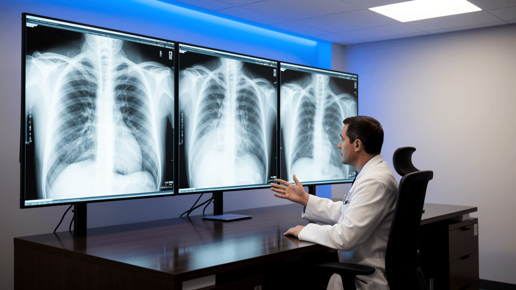
Emergency departments and intensive care units may soon gain access to faster, more portable AI tools for detecting pneumothorax—a potentially life-threatening condition where air leaks into the space between the lung and chest wall—thanks to a novel training technique that compresses diagnostic intelligence into models small enough to run on mobile devices.
Researchers Tao Hong and Hao Chen have developed a mask-aware knowledge distillation framework that enables a compact MobileNetV3-Small neural network to match the pneumothorax classification performance of a full-scale ResNet-50 model while requiring a fraction of the computational resources. Published in Medical and Biological Engineering and Computing, the study demonstrates how auxiliary lesion segmentation masks used exclusively during training can guide a lightweight "student" model to learn spatial attention patterns from a larger "teacher" network, eliminating the need for computationally expensive segmentation decoders during clinical use.
Matching Full-Scale Performance in a Pocket-Sized Package
Working with 10,675 chest radiographs from the SIIM-ACR Pneumothorax Segmentation dataset and 498 additional positive cases from the PTX-498 dataset, the research team trained their compact student model to achieve a test area under the curve (AUC) of 0.946—statistically indistinguishable from the teacher model's 0.950 AUC according to DeLong testing with Holm correction. The locked 20% hold-out test set comprised 2,135 radiographs, stratified and validated via SHA-256 file hashing.
The mask-aware approach proved critical: when compared to a uniform-attention control, the mask-guided student achieved significantly superior discrimination (AUC 0.946 vs. 0.936, paired DeLong p=0.003024). Ablation studies revealed that removing attention transfer from the deepest network layer reduced test AUC to 0.931, while eliminating logits distillation dropped performance to 0.934.
At operating points calibrated for clinical triage, the student ensemble achieved 92.9% sensitivity with 75.9% specificity at a validation-derived 90% sensitivity threshold. When tuned for maximum case-finding, the model reached 97.3% sensitivity, though specificity fell to 62.2%—a trade-off the researchers note would require radiologist confirmation to manage false-positive alerts in practice.
From Research Workstation to Bedside Device
The framework addresses a persistent bottleneck in emergency radiology: existing high-performance AI models typically require server-grade hardware that limits deployment to centralized PACS systems or cloud infrastructure. By achieving near-teacher-level discrimination with a model architecture designed for mobile processors, the approach opens pathways for real-time computer-aided diagnosis on portable X-ray machines, bedside workstations, and edge devices in resource-constrained settings.
Training employed standard protocols—50 epochs using AdamW optimization on an NVIDIA RTX 4090 GPU, with five-fold cross-validation yielding mean validation AUC of 0.938 (standard deviation 0.009) for both student and teacher models. Images were preprocessed to single-channel 8-bit grayscale at 512×512 pixel resolution, with augmentation including horizontal flips, ±15° rotation, and brightness perturbation. Both networks used ImageNet pretraining, and the distillation process incorporated dynamic annealing of temperature scaling and loss weights over the initial 60% of training.
Benchmark comparisons showed the student ensemble statistically outperformed EfficientNet-B0, hard-label-only training, vanilla distillation, and ablated variants, while showing no significant difference from DenseNet121.
Clinical Implications and Next Steps
The high-sensitivity operating points demonstrated in the study—particularly the 97.3% sensitivity threshold—align with triage workflows where missing a pneumothorax carries severe clinical consequences, including respiratory failure and cardiovascular collapse. However, the corresponding specificity limitations underscore that AI-assisted screening should complement rather than replace radiologist interpretation, especially given the life-threatening nature of missed diagnoses.
The reliance on publicly available datasets and standard pretrained architectures may lower barriers to replication and regulatory validation for companies developing point-of-care diagnostic tools or AI-enabled radiology integrations. The method's ability to retain spatial reasoning capabilities without deploying dual-branch segmentation decoders during inference represents a practical solution to the latency-accuracy trade-off that has historically constrained lightweight models in acute thoracic imaging.
For emergency departments and intensive care units, faster automated pneumothorax screening could reduce time-to-diagnosis when subtle radiographic findings might otherwise be overlooked or delayed pending specialist review—though prospective calibration of decision thresholds in clinical settings remains essential before deployment.
Sources
Follow Health AI Central
Keep up with health AI where you already read
On LinkedIn
The day’s strongest health AI stories in your feed, one or two each weekday.
On Google
Choose Health AI Central as a preferred source, and Google is more likely to show our reporting in Top Stories.
Reporting & accountability
How we maintain accuracy
Health AI Central separates reported facts, analysis, and public-source data. Readers can review our standards, request a correction, or consult the relevant research methodology before relying on a claim.