

Open-Source AI Uncovers Hidden Geometry That Predicts Kidney Tumor Treatment Failure
Taiwanese researchers demonstrate that automated measurement of tumor contact with renal sinus blood vessels outperforms traditional scoring systems in forecasting cryoablation outcomes.
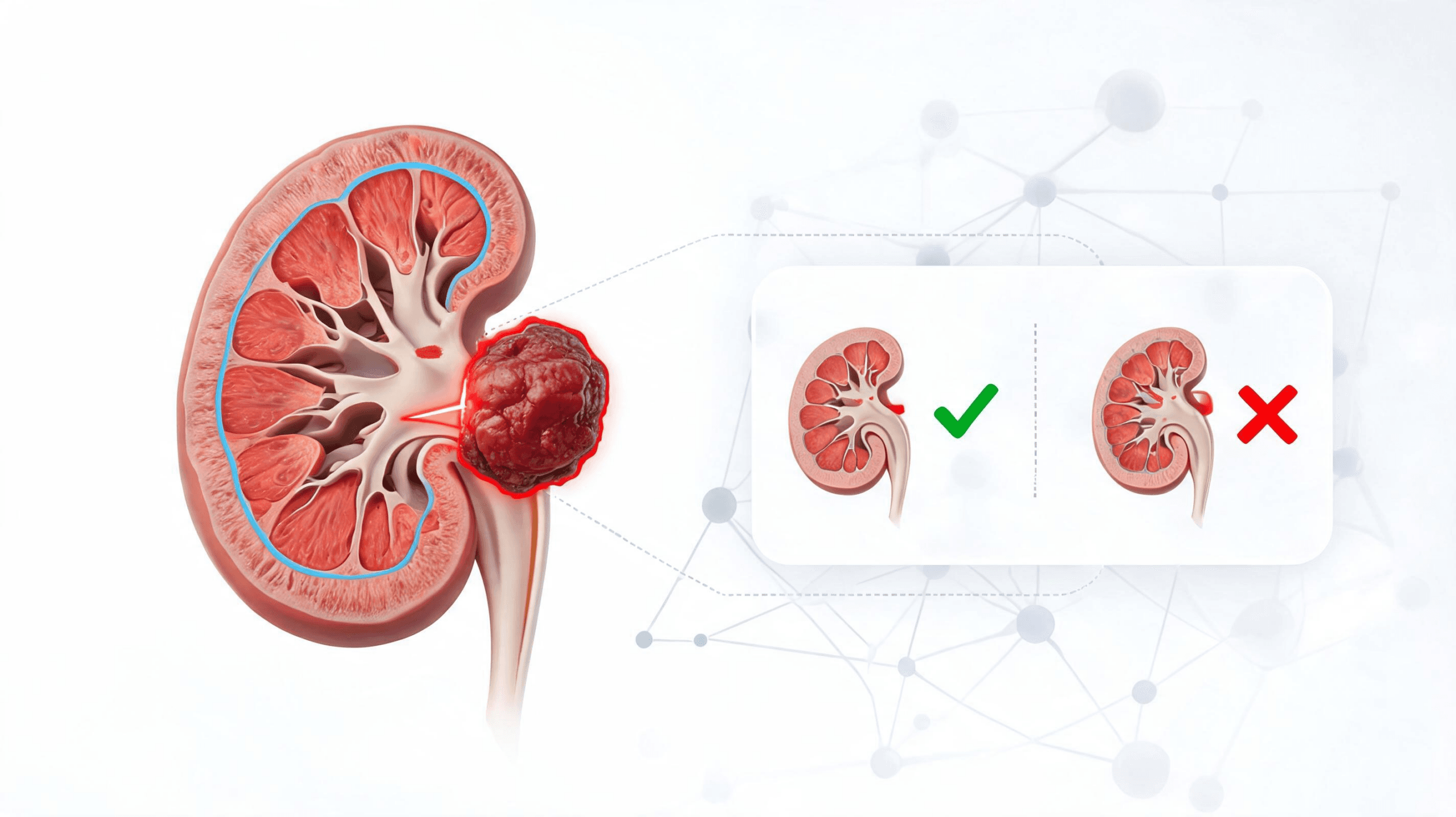
Interventional radiologists treating small kidney tumors may soon have a powerful new tool for predicting which patients face the highest risk of treatment failure—and it comes from repurposing open-source artificial intelligence models originally built for other purposes. Researchers at Taipei Veterans General Hospital have developed an automated workflow that measures a previously underappreciated three-dimensional feature: the contact area between tumors and the renal sinus, the kidney's central blood vessel-rich region.
Their findings, published in CVIR Oncology, suggest this AI-derived metric captures anatomical risk factors that conventional scoring systems miss entirely.
A Five-Minute Analysis That Outperforms Manual Scoring
The research team analyzed 116 patients who underwent CT-guided percutaneous cryoablation for renal lesions between October 2009 and December 2024, with mean follow-up of 2.8 years. Using two open-source deep learning models—TotalSegmentator and KiTS23, both built on the nnU-Net framework—they automatically segmented tumors and surrounding anatomy from pre-treatment CT scans in approximately five minutes per case.
The key finding: tumor-renal sinus contact area emerged as the only independent predictor of treatment failure in multivariable analysis, with an odds ratio of 1.379 (p=0.002). A composite risk score combining this contact area measurement with tumor volume achieved an area under the curve of 0.765 for predicting local tumor control failure, compared to 0.664 for the conventional RENAL nephrometry score.
Among the 116-patient cohort, 94% achieved complete ablation at first follow-up, but 6.9% of those with initially successful treatment later developed local tumor progression. The automated risk score, using an optimal cutoff of 1.51, identified high-risk cases with 67% sensitivity and 86% specificity. Cox survival analysis confirmed that larger tumor-renal sinus contact area significantly associated with progression (hazard ratio 1.255, p=0.026).
Why Blood Vessel Proximity Matters for Freezing Tumors
The researchers propose that extensive tumor contact with renal sinus structures creates a "cold-sink effect"—blood flow through nearby vessels dissipates the freezing energy needed to kill cancer cells, similar to the well-documented heat-sink phenomenon that limits radiofrequency ablation efficacy. While the study did not directly measure this mechanism, the clinical association proved robust across 15 years of procedural variations.
Notably, only 16 of 116 patients (13.8%) required any manual correction to the automated segmentation masks, and no cases needed complete re-segmentation. This high accuracy rate suggests the workflow could integrate into clinical practice without overwhelming radiologist workload.
The study illustrated the clinical significance with contrasting cases: a patient with 0.31 cm² contact area (risk score 0.59) remained disease-free for over four years, while another with 9.23 cm² contact area (risk score 3.54) experienced tumor progression at nine months.
From Research to Clinical Reality
The automated approach addresses a known weakness of manual nephrometry scoring: substantial interobserver variability, particularly in assessing tumor location and exophytic properties. By extracting continuous volumetric features from full three-dimensional anatomy rather than relying on measurements in orthogonal planes, the AI system captures spatial relationships that categorical scoring systems cannot represent.
For clinical deployment, the findings suggest opportunities to objectively identify high-risk cases during pre-procedural planning, potentially guiding treatment selection between ablation and surgery or tailoring post-procedure surveillance intensity. However, the researchers acknowledge important limitations: the retrospective single-center design, relatively small number of treatment failures (15 cases), and lack of external validation mean the tool requires testing in other institutions before routine clinical use.
The demonstration that open-source segmentation models can enable novel biomarker discovery without proprietary algorithm development may accelerate similar applications across interventional oncology, wherever vascular anatomy influences thermal ablation outcomes.
Sources
Google Search
Follow Health AI Central on Google
Choose Health AI Central as a preferred source, and Google is more likely to show our reporting in Top Stories.
Reporting & accountability
How we maintain accuracy
Health AI Central separates reported facts, analysis, and public-source data. Readers can review our standards, request a correction, or consult the relevant research methodology before relying on a claim.