
Your Face May Reveal Your Cancer Survival Odds Before Treatment Begins
Mass General Brigham researchers have developed an AI system that analyzes a single facial photograph to predict biological age and mortality risk in cancer patients, validated across more than 43,000 cases.
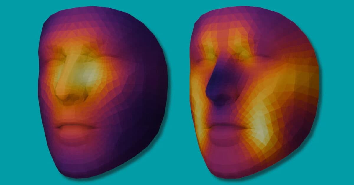
A simple photograph could soon help oncologists determine which cancer patients are most likely to survive intensive treatment, according to research from Mass General Brigham Cancer Institute. The team has developed FAHR-Face (Foundation Artificial Intelligence for Health Recognition), an AI foundation model that extracts health information from facial images to assess both biological aging and cancer treatment outcomes.
The technology represents a departure from traditional frailty assessments, which often require time-consuming physical tests that can be burdensome for already-compromised cancer patients. Instead, FAHR-Face analyzes consumer-grade photographs to generate risk stratification data within moments.
Two Tools, One Foundation Model
The FAHR-Face system spawned two specialized clinical applications: FAHR-FaceAge and FAHR-FaceSurvival. Both tools emerged from a foundation model trained on more than 42 million facial photographs using masked autoencoder self-supervised learning—a technique that allows the AI to learn patterns without explicit labeling.
FAHR-FaceAge estimates biological age with a mean absolute error of just 5.1 years, outperforming competing models that averaged 6.92 years of error. The tool focuses on specific anatomical regions including nasolabial folds, the forehead above the orbits, and the temples. According to the research published in The Lancet Digital Health, patients who appeared five or more years older than their chronological age faced 21% higher mortality risk, while those appearing five years younger enjoyed 17% higher survival rates.
FAHR-FaceSurvival directly predicts cancer treatment outcomes. Trained on more than 34,000 cancer patients undergoing radiotherapy and validated on two additional cohorts totaling 9,000 patients, this tool evaluates the region below the eyes and nasal bridge to stratify mortality risk.
Dramatic Risk Stratification
The survival prediction tool demonstrated striking accuracy in identifying high-risk patients. Those classified in the highest-risk category experienced mortality rates three times greater than lowest-risk patients. Perhaps most significantly, 52% of highest-risk patients died within 90 days compared to just 2.1% in the lowest-risk group—a difference that could fundamentally alter treatment decision-making.
Dr. Raymond Mak, a radiation oncologist and corresponding author on the study, and Dr. Hugo Aerts, co-senior author and Director of the AI in Medicine Program at Mass General Brigham, led the research demonstrating the system's robustness across diverse conditions. The model proved unaffected by makeup, facelifts, glasses, or lighting variations, though it showed minor errors with rhinoplasty patients.
Implications for Cancer Care
For healthcare providers, FAHR-Face offers a rapid, non-invasive screening tool to supplement clinical judgment when assessing whether elderly patients can tolerate intensive interventions like radiation, surgery, or systemic therapy. The technology could enable earlier, more informed conversations about treatment intensity and goals of care based on objective biological markers rather than chronological age alone.
The foundation model approach also signals a methodological shift in medical AI development. Rather than building separate task-specific models from scratch, researchers can now fine-tune a single large-scale pre-trained system for multiple clinical applications—potentially accelerating development of facial biomarker tools across conditions beyond oncology.
Mass General Brigham has made a 10-minute study available online for public participation in FaceAge assessment, suggesting the team continues to refine and expand the technology's capabilities.
Sources
Google Search
Follow Health AI Central on Google
Choose Health AI Central as a preferred source, and Google is more likely to show our reporting in Top Stories.
Reporting & accountability
How we maintain accuracy
Health AI Central separates reported facts, analysis, and public-source data. Readers can review our standards, request a correction, or consult the relevant research methodology before relying on a claim.