

Tiny Biopsy Samples, Big AI Predictions: New Tool Could Spare 95% of Gastric Cancer Patients from Unnecessary Surgery
Shanghai researchers demonstrate that artificial intelligence can predict lymph node spread in early gastric cancer using only 2-millimeter endoscopic biopsies, potentially eliminating the need for radical stomach removal in the vast majority of low-risk patients.
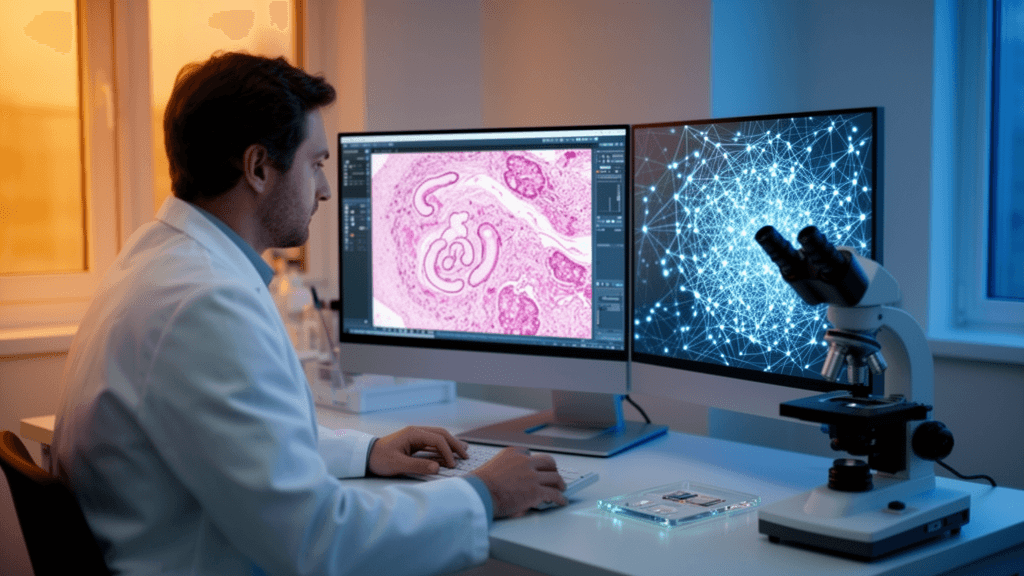
A multimodal artificial intelligence framework developed at Zhongshan Hospital of Fudan University in Shanghai is demonstrating the potential to transform treatment decisions for early gastric cancer by predicting lymph node metastasis risk from preoperative endoscopic biopsy slides—tissue samples barely 2 millimeters across that are available at initial diagnosis rather than after surgical resection.
The system addresses a critical clinical dilemma: current risk stratification tools frequently trigger radical gastrectomy in patients who have undergone endoscopic resection, yet 90–95% of these follow-up surgeries find no metastasis, exposing patients to unnecessary surgical morbidity including anastomotic leak, dumping syndrome, and nutritional deficiency. Gastric cancer remains the fourth most prevalent malignancy worldwide and the third leading cause of cancer death, with East Asia accounting for more than 70% of the global burden.
Extracting Signal from Fragmented Specimens
The research team analyzed 637 patients with pathologically confirmed T1 gastric cancer treated between January 2008 and December 2022, processing 1,099 surgical whole slide images and 189 paired endoscopic biopsy slides. The AI framework achieved an area under the curve (AUC) of 0.759 on surgical tissues and 0.727 on preoperative endoscopic biopsy slides—performance that represents measurable progress on one of computational pathology's most challenging problems.
The technical architecture employs a dual-stream Transformer with a clinical-guided co-attention mechanism, leveraging the Prov-GigaPath foundation model, a Vision Transformer Giant encoder with 1.3 billion parameters trained on 1.3 billion image tiles. External validation on the TCGA-STAD cohort yielded an AUC of 0.751 (95% CI 0.613–0.892).
At the study's 13.2% metastasis prevalence, the biopsy model achieved sensitivity of 0.733, specificity of 0.682, projected positive predictive value of 26.0%, and negative predictive value of 94.4%. The high negative predictive value suggests the tool functions best as a rule-out test, identifying low-risk patients who could proceed directly to organ-preserving endoscopic resection.
Curriculum Learning Bridges the Data Gap
A key innovation involves two-stage curriculum learning: the system was first trained on spatially contiguous "pseudo-biopsies" extracted from surgical specimens using unsupervised segmentation, then fine-tuned on fragmented real endoscopic biopsy slides. This strategy addresses the scarcity of labeled preoperative specimens while teaching the model to handle the spatial fragmentation inherent in endoscopic biopsies.
The automated segmentation pipeline—using t-SNE projection, K-means clustering, and quality control—achieved a mean Dice coefficient of 0.846 and mean precision of 0.926 against expert annotations. Expert pathologists rated 89.6% of the 1,099 slides as excellent quality, with overall clinical usability reaching 94.5%.
Ablation studies confirmed the importance of each architectural component: removing the multiple-instance-learning Transformer dropped biopsy AUC to 0.612, omitting curriculum learning reduced performance to 0.635, and eliminating co-attention lowered it to 0.686. The framework outperformed established baselines by 3.1 percentage points over the strongest comparator.
Biological Validation and Clinical Gaps
Model attention maps concentrated on tumor tissue and tumor-infiltrating lymphocytes, with high-attention regions showing significantly greater tertiary lymphoid structure density (Wilcoxon p=0.0017). Tertiary lymphoid structure density in these regions was significantly higher in metastasis-positive versus metastasis-negative cases (Mann-Whitney U p<0.0001), suggesting the AI identifies biologically meaningful tissue patterns.
However, the authors specify that clinical deployment requires sensitivity of 90–95% and negative predictive value above 95%—thresholds not yet met. The current 26% positive predictive value means that 74 of 100 patients flagged as high-risk would not actually have metastasis, indicating the tool cannot yet autonomously guide treatment decisions without confirmatory assessment.
The research, published in Annals of Biomedical Engineering (DOI: 10.1007/s10439-026-04380-7), represents progress toward preoperative treatment personalization that could preserve quality of life and digestive function for thousands of gastric cancer patients annually, particularly in high-incidence regions, while maintaining oncologic safety. Prospective multi-institutional validation with calibration and net benefit assessment remains essential before clinical translation.
Sources
Reporting & accountability
How we maintain accuracy
Health AI Central separates reported facts, analysis, and public-source data. Readers can review our standards, request a correction, or consult the relevant research methodology before relying on a claim.