

Routine CT Scans Could Reveal Hidden Osteoporosis Risk Without Extra Radiation, Chinese Study Finds
Deep learning extracts bone aging patterns from standard imaging, outperforming chronological age in predicting bone density decline across 4,152 patients.
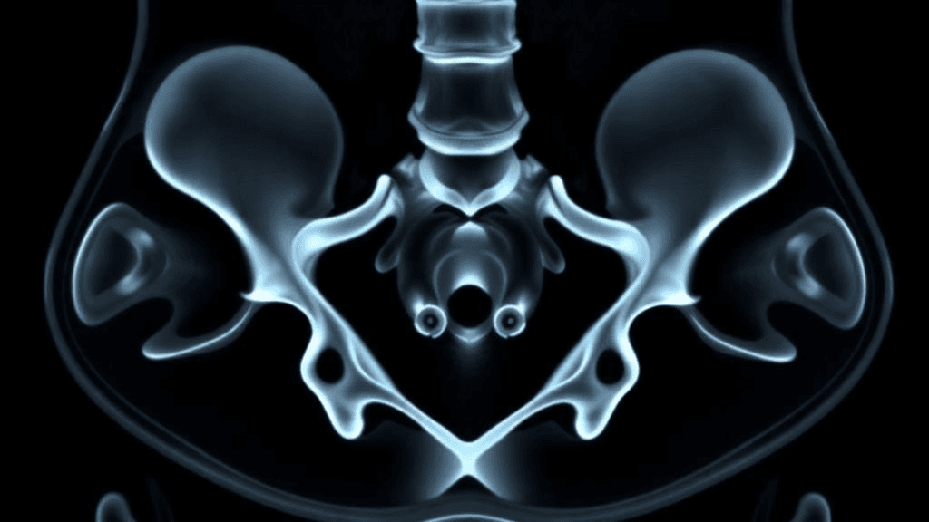
Millions of patients undergo CT scans each year for reasons unrelated to bone health—yet those same images contain untapped information about skeletal aging and fracture risk. A nationwide Chinese study published in BMC Medical Imaging now demonstrates that artificial intelligence can automatically read these hidden patterns, potentially transforming routine imaging into an opportunistic screening tool for osteoporosis without requiring additional scans or radiation exposure.
The retrospective multicohort study, registered as ClinicalTrials.gov NCT07162168 and led by researchers at Peking University People's Hospital, analyzed CT scans from 4,152 participants across Chinese hospitals. Using deep learning to segment the proximal femur and extract radiomic features, the team developed an age estimation model that correlated more strongly with actual bone mineral density than patients' chronological age alone.
AI Outperforms Calendar Age in Predicting Bone Density
The research team, led by Hanwen Cheng, Liwei Zhuang, and Yiran Zhang, employed an nnU-Net-based deep learning system to automatically segment the proximal femur from routine CT images, achieving a Dice similarity coefficient of 0.963—indicating near-perfect anatomical delineation. From these segmented regions, the PyRadiomics platform extracted standardized structural features that were fed into support vector regression models.
In the external validation cohort of 1,183 patients, the AI-derived radiomic age index showed a Spearman correlation of −0.546 with femoral neck volumetric bone mineral density, compared to −0.443 for chronological age (Steiger's Z = 4.97, P < 0.001). The system estimated chronological age with a mean absolute error of 8.84 years and R² of 0.50, while sex classification models achieved an area under the curve of 0.94 using both support vector classification and XGBoost algorithms.
The study population included 1,947 men with a median age of 59 years, spanning ages 18 to 99. The development cohort comprised 2,969 participants, with findings validated in the separate external cohort.
From Research Finding to Clinical Screening Tool
The stronger correlation between radiomic features and bone density suggests that structural patterns captured by AI may reflect biological aging processes more accurately than years lived. This finding supports the "opportunistic imaging" paradigm—leveraging scans already performed for abdominal, chest, or pelvic indications to identify osteoporosis risk in patients who might never receive dedicated bone density testing.
Current osteoporosis screening relies primarily on dual-energy X-ray absorptiometry (DXA), which remains chronically underutilized. By embedding bone health analytics into existing radiology workflows, the approach could catch high-risk patients before their first fracture without requiring additional imaging appointments or radiation dose.
However, the 8.84-year mean absolute error limits precision for individual patient age estimation, and the authors acknowledge that scanner variability, reconstruction protocol differences, and the single-population training cohort (Chinese patients) constrain immediate generalizability. Critically, the study correlated radiomic features with bone density but did not validate fracture prediction—the ultimate clinical endpoint required before deployment as a diagnostic tool.
What Comes Next
The research, funded by the Beijing Natural Science Foundation-Fengtai Joint Innovation Project, National Natural Science Foundation of China Science Center Program, and Shenzhen Medical Research Fund, was approved by the Peking University People's Hospital ethics committee with waived informed consent for retrospective de-identified images.
For healthcare AI companies developing bone health analytics, the study validates both the technical feasibility of automated skeletal radiomics and the potential to leverage existing CT infrastructure rather than requiring dedicated imaging protocols. The nnU-Net segmentation benchmark and multicohort validation design establish reproducibility standards for future commercial products.
Prospective studies linking radiomic bone age to fracture outcomes will determine whether this opportunistic screening approach can translate correlation into clinical utility—and whether hidden patterns in routine scans can finally close the osteoporosis detection gap.
Sources
Reporting & accountability
How we maintain accuracy
Health AI Central separates reported facts, analysis, and public-source data. Readers can review our standards, request a correction, or consult the relevant research methodology before relying on a claim.