

Pregnancy's Hidden Fracture Risk: How AI X-Ray Analysis Uncovered a Bone-Thinning Side Effect of Common Blood Thinner
University of Tokyo researchers used artificial intelligence to reveal that heparin therapy during pregnancy significantly reduces maternal bone density—a danger previously invisible because standard bone scans are avoided in expectant mothers.
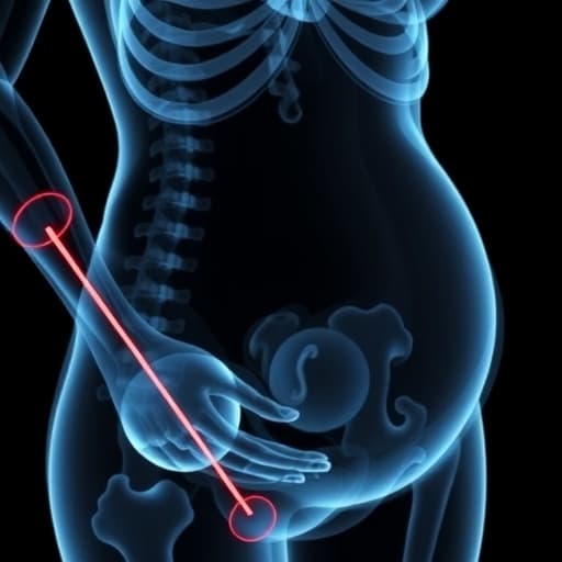
Pregnant women receiving a widely used blood thinner face a previously unquantified skeletal threat that has remained hidden because the gold-standard test for bone health is routinely avoided during pregnancy. Now, researchers at the University of Tokyo have deployed an AI system that converts ordinary chest X-rays into bone density assessments, revealing that unfractionated heparin significantly reduces maternal bone strength—particularly in underweight women—at the precise moment when the developing fetus demands massive calcium transfers from the maternal skeleton.
The findings, published in Reproductive Sciences, emerge from a collaboration between the University of Tokyo's departments of obstetrics, orthopedic surgery, and preventive medicine, along with technology partner KYOCERA Corporation. The study analyzed preoperative chest X-rays from 86 pregnant women who received heparin therapy compared to 213 controls undergoing cesarean delivery without anticoagulation, spanning April 2013 through October 2023.
AI Screening Where Standard Tests Cannot Go
The deep learning platform at the heart of this discovery was trained by pairing plain radiographs with conventional dual-energy X-ray absorptiometry (DXA) measurements—the clinical gold standard for bone density assessment. Once trained, the AI could estimate bone mineral density (eBMD) from chest X-rays alone, without the additional radiation exposure that makes DXA testing problematic during pregnancy.
This capability proved critical for detecting a hidden maternal health risk. Venous thromboembolism ranks among the leading causes of maternal death in high-income countries, and pregnancy naturally elevates clotting risk through venous stasis, vessel injury, and elevated clotting factors. Heparin therapy becomes medically necessary for conditions including antiphospholipid syndrome, inherited thrombophilias, mechanical heart valves, acute clots, severe kidney impairment, and urgent procedures requiring rapid anticoagulant reversal.
The AI analysis revealed that heparin-exposed women showed significantly lower estimated bone mineral density at both the lumbar spine and femoral neck. In multivariate analysis adjusting for body mass index and other covariates, unfractionated heparin exposure remained an independent risk factor for reduced lumbar spine bone density.
Underweight Women Face Greatest Skeletal Vulnerability
The skeletal impact proved most severe in underweight women, defined by Asian BMI thresholds. This subgroup demonstrated significantly lower bone density, with the deficit most pronounced when low body weight combined with heparin exposure. The lumbar spine—rich in trabecular bone—showed greater vulnerability than the femoral neck, which contains more cortical bone.
Paradoxically, researchers found no significant correlation between cumulative heparin dose and bone density reduction, suggesting the relationship may be more complex than simple dose-response. The mechanisms behind heparin's bone toxicity include suppression of osteoblasts (bone-building cells), promotion of osteoclast-driven resorption, calcium binding, and disruption of vitamin D metabolism—all occurring as the fetus accumulates approximately 30 grams of calcium during pregnancy, predominantly in the third trimester.
From Research Tool to Clinical Surveillance
The AI platform had been previously described in the Journal of Orthopaedic Research and validated in postmenopausal women after hip replacement. Its application to pregnancy represents a novel use case: opportunistic screening from images already obtained for surgical clearance, enabling bone health surveillance in a population where pregnancy-associated osteoporosis often goes undetected until devastating vertebral fragility fractures occur in late pregnancy or lactation—events frequently dismissed initially as ordinary back pain.
For clinicians managing high-risk pregnancies requiring anticoagulation, the findings suggest potential strategies including preferential use of low-molecular-weight heparin when medically appropriate, heightened monitoring of underweight patients on unfractionated heparin, and calcium and vitamin D supplementation. The AI technology offers a method to quantify skeletal risk using imaging already acquired for other clinical indications.
The research received funding from Japan's Ministry of Health, Labour and Welfare, the Japan Society for the Promotion of Science, and KYOCERA Corporation. The technology's ability to extract quantitative bone data from routine radiographs positions it as a scalable screening model with minimal workflow disruption—addressing a clinical blind spot in the millions of annual births globally where pregnant women require anticoagulation therapy.
Sources
Reporting & accountability
How we maintain accuracy
Health AI Central separates reported facts, analysis, and public-source data. Readers can review our standards, request a correction, or consult the relevant research methodology before relying on a claim.