

Machine Learning Unveils Invisible Brain Damage in Newborns That Radiologists Miss
A new AI model detects hypoxic-ischemic encephalopathy in infants with normal-appearing MRI scans, achieving 87.8% accuracy in prospective validation and potentially preventing lifelong developmental consequences.
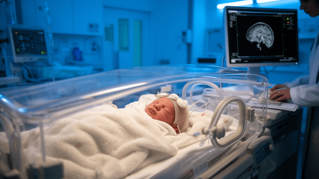
When a newborn suffers oxygen deprivation during birth, the resulting brain injury can be devastating—yet conventional MRI scans often appear completely normal to the trained eye. Now, researchers at Children's Hospital of Soochow University have developed a machine learning tool that identifies hidden hypoxic-ischemic encephalopathy (HIE) in infants whose scans look reassuringly clear to radiologists.
The breakthrough addresses what clinicians call the "visually normal MRI paradox": babies with birth asphyxia who appear to have escaped brain injury on standard imaging, yet go on to develop significant neurodevelopmental problems. For these infants, the window for early intervention quietly closes while families receive false reassurance.
Radiomics Reveals Subtle Patterns Invisible to Human Eyes
The research team, led by Yong-hao Yue and colleagues, analyzed MRI scans from 297 term newborns with birth asphyxia treated between January 2020 and December 2025, then prospectively validated their model on 54 additional infants between January and April 2026. Published in BMC Medical Imaging, the study used radiomics—a technique that extracts hundreds of quantitative features from medical images that are imperceptible to human observers.
The algorithm focused on two brain regions particularly vulnerable to oxygen deprivation: the basal ganglia and thalami. By analyzing T1-weighted, T2-weighted, and diffusion-weighted MRI sequences, the model identified six sequence- and region-specific radiomics scores that, when combined with clinical data, achieved an area under the curve (AUC) of 0.958 in the training set, 0.915 in the test set, and 0.878 in prospective validation.
The combined clinical-radiomics model significantly outperformed all comparator approaches, including clinical assessment alone and radiomics-only models, according to DeLong test analysis. SHAP (SHapley Additive exPlanations) analysis revealed that T2-weighted and diffusion-weighted imaging features carried the highest predictive importance, providing clinician-facing explanations that could facilitate regulatory approval and clinical adoption.
Clinical Utility Demonstrated Across Multiple Sites
Decision curve analysis confirmed the model delivers net clinical benefit compared to either treating all at-risk infants aggressively or treating none—the current extremes that clinicians navigate when faced with diagnostic uncertainty. The tool was validated across two sites: Children's Hospital of Soochow University and Yulin Children's Hospital, demonstrating generalizability beyond a single institution.
For neonatal intensive care units, this represents a potential shift in risk stratification. Infants with undetected HIE may miss critical windows for neuroprotective interventions and early developmental support, potentially affecting lifelong cognitive and motor outcomes. An objective tool identifying hidden injury could reduce under-triage, ensure appropriate follow-up, and provide families with earlier, more accurate prognostic information.
The research, funded by the Natural Science Foundation of Jiangsu Province, the National Natural Science Foundation of China, and the Suzhou Medical Key Discipline program, establishes a blueprint for multimodal AI tools in pediatric radiology—combining quantitative imaging biomarkers with clinical data rather than relying on imaging alone.
As neonatal AI moves from laboratory validation to clinical implementation, this study demonstrates that machine learning can augment clinical judgment in high-stakes diagnostic scenarios where human perception reaches its limits.
Sources
Reporting & accountability
How we maintain accuracy
Health AI Central separates reported facts, analysis, and public-source data. Readers can review our standards, request a correction, or consult the relevant research methodology before relying on a claim.