

Blood Cancer Detection Breakthrough: New AI Architecture Fuses Local and Global Vision to Achieve Near-Perfect Accuracy
Researchers combine mobile convolutional networks with vision transformers and adaptive attention to distinguish leukemia cells from healthy look-alikes in microscopic blood images.
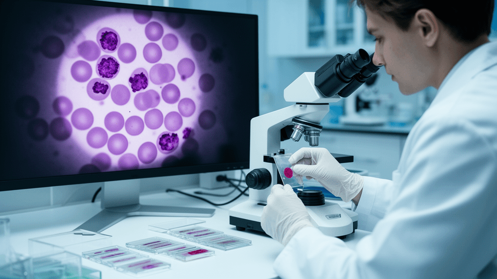
Distinguishing cancerous blast cells from their healthy mimics in blood smears has long challenged pathologists screening for acute lymphoblastic leukemia (ALL), particularly when differentiating malignant cells from normal hematogones that share similar morphological features. Now, researchers at India's Vellore Institute of Technology report a hybrid artificial intelligence model that achieved 99% accuracy on a benchmark dataset by strategically combining two complementary computer vision paradigms—convolutional neural networks for fine morphological detail and vision transformers for global contextual relationships.
Dual-Vision Architecture Targets Diagnostic Blind Spot
The model, dubbed MoViTHAFX-Net, addresses a fundamental tension in medical image analysis: convolutional neural networks excel at capturing local features like nuclear texture and cytoplasmic granularity, while vision transformers process entire image fields to understand spatial relationships and contextual patterns. Rather than choosing one approach, researchers A. Elavarasi and N. Jaisankar engineered a system that adaptively weights both streams using a novel Hierarchical Attention Fusion mechanism.
Working with the C-NMC 2019 benchmark dataset containing 10,661 labeled peripheral blood smear images, the team split data into 7,462 training, 1,600 validation, and 1,599 test images using image-level stratified partitioning. On the held-out test set, MoViTHAFX-Net correctly classified 1,583 of 1,599 images, achieving 99.00% accuracy alongside precision, recall, and F1-scores of approximately 0.990 and an area under the curve of 0.99. The architecture pairs MobileNet—a lightweight convolutional network designed for efficiency—with a Vision Transformer backbone, integrating their outputs through the hierarchical attention layer that learns which feature stream to prioritize for each diagnostic decision.
Interpretability and Clinical Translation Challenges
To address the "black box" criticism often leveled at deep learning diagnostics, the researchers incorporated Grad-CAM++ visualization, which generates heatmaps highlighting image regions that drive each classification. These attention maps typically focus on nuclear morphology and other clinically relevant features, offering pathologists insight into the model's reasoning process—a critical requirement for clinical trust and regulatory acceptance.
However, the researchers explicitly acknowledge significant limitations that separate benchmark performance from clinical readiness. The image-level data split means the same patient's cells could appear in both training and test sets, potentially inflating performance estimates. More fundamentally, validation on a single dataset provides no evidence of generalization across diverse patient populations, staining protocols, imaging equipment, or healthcare settings—the real-world variability that determines whether 99% benchmark accuracy translates to reliable clinical performance.
From Research Milestone to Diagnostic Tool
The work, published in Scientific Reports with no external funding, represents a technical proof-of-concept rather than a deployable diagnostic system. Ablation studies showed the Hierarchical Attention Fusion mechanism provided incremental advantages over simple feature concatenation within the experimental framework, suggesting the adaptive weighting strategy captures complementary information from both vision paradigms.
For the technology to progress from research milestone to clinical tool, future validation must employ subject-disjoint data splits, multi-center testing across varied imaging conditions, and prospective studies comparing AI-assisted versus unassisted pathologist performance in real diagnostic workflows. If such validation succeeds, the hybrid architecture could reduce diagnostic variability and pathologist fatigue in ALL screening, particularly benefiting resource-limited settings where access to experienced hematopathologists remains constrained.
Sources
Reporting & accountability
How we maintain accuracy
Health AI Central separates reported facts, analysis, and public-source data. Readers can review our standards, request a correction, or consult the relevant research methodology before relying on a claim.