

Machine Learning Cracks the Code on Hard-to-Diagnose Digestive Tumors Using Routine Hospital Data
Chinese researchers achieve 96% accuracy identifying gastrointestinal stromal tumors by fusing CT imaging features with standard blood tests, potentially eliminating risky biopsies.
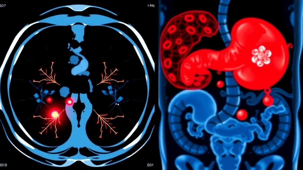
Gastrointestinal stromal tumors occupy a diagnostic gray zone that has long frustrated oncologists: too rare to screen for systematically, too dangerous to miss, and risky to biopsy. Now, a team from Gansu Provincial Hospital and collaborating institutions has demonstrated that machine learning can identify these elusive mesenchymal tumors before surgery with near-pathological certainty—using only data already collected in routine clinical workflows.
The researchers developed a system that extracts quantitative features from standard contrast-enhanced CT scans and combines them with five blood biomarkers, achieving an area under the ROC curve of 0.96 on independent testing. Published in Holistic Integrative Oncology, the study analyzed 317 patients treated between January 2017 and December 2024, comparing 115 GIST cases against 202 controls with other digestive diseases including gastric cancer, colorectal cancer, and benign conditions.
Fusing Imaging Physics with Blood Chemistry
The breakthrough lies in multimodal integration. Lead researchers Jing Yang and Hui Cai, working with teams from Southern Medical University and Gansu University of Chinese Medicine, started with 851 radiomic features extracted from portal-phase CT scans—quantitative descriptors of tumor shape, texture, and intensity patterns. LASSO regression winnowed these to four critical features: Sphericity, RunVariance, SurfaceVolumeRatio, and LongRunEmphasis.
From a panel of 42 fasting blood indicators spanning hematology, biochemistry, and tumor markers, five emerged as independent predictors: direct bilirubin, alkaline phosphatase, CEA, CA199, and the pepsinogen I/II ratio. GIST patients exhibited a distinctive signature—higher bilirubin and pepsinogen ratios but lower CEA and CA199 compared to epithelial cancers, reflecting their different cellular origin.
Testing six machine learning architectures, the team found LightGBM delivered optimal performance. Blood tests alone achieved only 0.745 AUC, confirming that laboratory values are insufficient without imaging context. Adding the four radiomic features increased accuracy to 0.964, demonstrating that imaging AI drives the diagnostic value.
Interpretability Meets Clinical Reality
Addressing the black-box criticism that shadows medical AI, the researchers deployed SHAP analysis to quantify each feature's contribution. Sphericity emerged as the dominant predictor with a mean absolute SHAP value of 2.68, followed by SurfaceVolumeRatio, while direct bilirubin contributed least. Decision curve analysis showed that at a 0.4 probability threshold, the model would yield approximately 35 additional net-benefit cases per 100 patients compared to a treat-none strategy.
The clinical stakes are substantial: GIST represents 0.1–3% of gastrointestinal malignancies, yet approximately 50% of patients already have metastases at diagnosis, and recurrence risk remains high even after resection. Current practice often requires invasive biopsy, which carries complication risks including theoretical tumor seeding. A non-invasive preoperative diagnostic tool could accelerate surgical planning and catch tumors in the pre-metastatic window.
Limitations and the Path Forward
The study's single-center retrospective design and small independent test set of 32 patients represent significant caveats. More notably, the control group excluded similar mesenchymal tumors such as leiomyoma and schwannoma—the very differential diagnoses that challenge radiologists in practice. Decision tree and LightGBM models achieved perfect 1.000 AUC on training data, signaling potential overfitting despite validation protocols.
The researchers relied on manual tumor segmentation by two radiologists with at least five years' experience, with senior arbitration for discrepancies, using an interclass correlation coefficient threshold of 0.75. Images were resampled to 1 mm³ voxels for standardization. Patients ranged from 29 to 81 years old, with a mean age around 60, and were split 7:2:1 across training, validation, and test sets.
Commercial and Clinical Implications
For health AI developers, the architecture validates a radiomics-plus-biomarker fusion approach that could integrate into existing PACS or radiology reporting systems without requiring specialized imaging protocols. The reliance on portal-phase CT—already standard in abdominal workups—broadens the addressable market compared to techniques requiring novel sequences.
For clinicians, the system offers a potential pathway to reduce diagnostic uncertainty and biopsy complications, though multicenter validation in populations including the full spectrum of mesenchymal tumor mimics remains essential before clinical deployment. The explicit use of interpretability methods addresses regulatory and trust requirements that have slowed medical AI adoption.
The study demonstrates that when machine learning is pointed at the right combination of routine data streams, even rare and diagnostically challenging cancers may yield their secrets—no new tests required.
Sources
Reporting & accountability
How we maintain accuracy
Health AI Central separates reported facts, analysis, and public-source data. Readers can review our standards, request a correction, or consult the relevant research methodology before relying on a claim.