

How CT Scanners Are Training X-Ray AI to Diagnose Lung Disease in Data-Starved Clinics
A new knowledge distillation framework transfers CT expertise to chest X-ray models, boosting accuracy by 10 percentage points with as few as 500 training images.
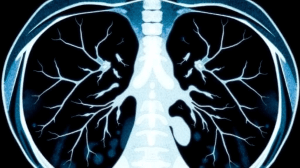
When medical AI needs thousands of labeled images to learn, clinics in resource-limited settings face an impossible barrier. But researchers at Manipal University Jaipur have found an ingenious workaround: teaching chest X-ray algorithms using the superior diagnostic knowledge embedded in CT scan models—without requiring paired imaging from the same patients.
Their CTGuideNet framework, published in Discover Artificial Intelligence, demonstrates that cross-modality "knowledge distillation" can achieve dramatic performance gains when labeled X-ray data is scarce, potentially bringing advanced lung disease screening to facilities where CT scanners remain out of reach.
Transferring Expertise Across Imaging Modalities
The research team—Preeti Sharma, Govind Murari Upadhyay, and Devershi Pallavi Bhatt—first trained a "teacher" network on 8,408 lung CT images spanning COVID-19, pneumonia, lung tumors, and normal tissue. This teacher achieved 98.10% accuracy with perfect precision and recall for COVID-19 detection and 100% recall for pneumonia.
The breakthrough came in transferring this CT-level expertise to "student" networks analyzing chest X-rays. Using a three-component loss function combining classification, normalized feature distillation, and spatial attention transfer, the framework guided X-ray models without requiring logit-level distillation or matched patient scans.
When trained on just 500 chest X-rays—100 per disease category—CT-guided supervision delivered accuracy improvements up to 10 percentage points. ResNet50 jumped from 71% to 81% accuracy, VGG16 climbed from 66% to 73%, and EfficientNet-B0 reached 82%. For lung tumor detection specifically, ResNet50 sensitivity surged from 50% to 83.33% with CT guidance.
Architecture Matters: One Size Does Not Fit All
A critical finding emerged during optimization: different neural network architectures required dramatically different distillation strategies. Feature distillation and attention transfer only improved performance when combined with architecture-aware weighting—each component alone actually underperformed baseline models.
VGG16 and MobileNetV2 tolerated strong feature and attention guidance, while ResNet variants needed weaker adaptive supervision. EfficientNet-B0 demanded a semantic-adaptive strategy with minimal cosine-based supervision at a coefficient of just 0.0005. This architecture-specific tuning proved essential for reliable cross-modality knowledge transfer.
External validation on the JSRT lung nodule dataset—247 Japanese chest radiographs—confirmed real-world applicability. Guided MobileNetV2 reached 86% accuracy versus 78% baseline, with F1-scores rising from 56% to 77%. Non-nodule recall improved from 16% to 50% while maintaining over 97% nodule sensitivity. Statistical significance was confirmed through paired t-tests and Wilcoxon signed-rank tests across eight random seeds at p < 0.05.
Implications for Resource-Limited Healthcare
The framework's performance at scale remained impressive. With 10,000 X-rays, EfficientNet-B0 with CT guidance achieved 94.60% accuracy across five disease categories including bacterial pneumonia, viral pneumonia, COVID-19, lung tumors, and normal tissue.
For healthcare providers, this approach enables accurate lung disease screening in settings where CT scanners are unavailable but chest X-rays remain routine. The technology could improve early detection without requiring expensive imaging infrastructure or large labeled datasets—critical advantages for under-resourced clinics.
For health AI companies, the cross-modality distillation methodology offers a pathway to build high-performing diagnostic models when labeled training data is scarce or expensive. The architecture-aware tuning blueprint provides guidance for optimizing knowledge transfer across different neural network families deployed on varied hardware from cloud servers to edge devices.
Patients in low-resource settings stand to benefit from more accurate X-ray interpretation, potentially reducing missed diagnoses and unnecessary CT referrals. The dramatic improvement in early-stage lung tumor sensitivity—from 50% to 83%—could enable earlier intervention when treatment outcomes are most favorable.
Sources
Reporting & accountability
How we maintain accuracy
Health AI Central separates reported facts, analysis, and public-source data. Readers can review our standards, request a correction, or consult the relevant research methodology before relying on a claim.