

Hidden Jawbone Infections Meet Their Match: New AI Sees Through Dental Anatomy to Detect What Clinicians Miss
A multi-task deep learning system achieves near-perfect discrimination of apical periodontitis on routine X-rays by learning to navigate the anatomical clutter that confounds human observers.
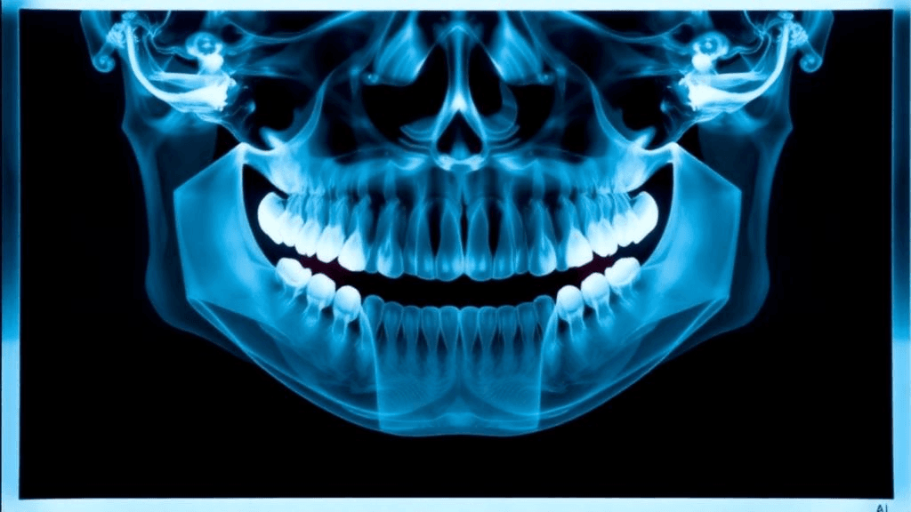
Periapical dental X-rays are among the most frequently captured medical images worldwide, yet the inflammatory bone lesions they're meant to reveal often hide in plain sight—obscured by overlapping nasal cavities, sinus floors, and mandibular bone. Now, a research collaboration spanning Fujian Medical University, the University of Hong Kong, and Tongji University has unveiled ApexTransNet, a deep learning framework that detects apical periodontitis—bacterial infections at tooth root tips—with 80.2% precision and near-perfect case-level discrimination, published in BMC Medical Imaging.
The system's innovation lies not in raw detection power alone, but in how it mimics the multi-layered reasoning of expert radiologists: simultaneously segmenting lesions, pinpointing their location, classifying disease presence, and refining boundaries—all while using anatomy-guided attention mechanisms to filter out the structural noise that trips up both algorithms and clinicians.
Anatomy-Aware Architecture Tackles Clinical Realism
ApexTransNet builds on a ResNet34 encoder backbone but adds two critical innovations. First, a Transformer-ASPP module combines atrous spatial pyramid pooling with Transformer attention layers, allowing the network to capture context at multiple scales while focusing computational resources on diagnostically relevant regions. Second, an anatomy-guided fusion stage explicitly teaches the model to distinguish pathological radiolucency from normal anatomical voids like the maxillary sinus.
Trained on 420 periapical radiographs from 600 unique patients—400 positive for apical periodontitis, 200 normal controls—the system was validated using patient-disjoint splits to prevent data leakage. Each positive case carried four independent expert annotations, reflecting the high inter-observer variability that plagues this diagnostic task in practice. On a 90-image test set, ApexTransNet achieved a Dice coefficient of 0.702, intersection-over-union of 0.575, and recall of 70.4%, with results stable across three random training seeds.
Near-Perfect Case Discrimination, But External Validation Required
At the case level—the clinically decisive question of "disease present or absent"—ApexTransNet delivered a ROC-AUC of 0.988 (95% CI: 0.965–0.999) and average precision of 0.995, with a Brier score of 0.067 indicating well-calibrated probability estimates. The system's 80.2% precision means that when it flags a lesion, it's correct four times out of five—a threshold designed to minimize false positives that could trigger unnecessary root canal procedures.
Yet the authors are explicit about constraints. The dataset came from a single health system with enriched disease prevalence, not representative of general clinic case mix. The precision-recall trade-off means ApexTransNet catches fewer true positives than some baseline models optimized purely for sensitivity. Most critically, the team emphasizes that external validation across diverse imaging equipment, patient populations, and clinical workflows is mandatory before any diagnostic claims.
Transparent AI for a High-Volume, High-Variability Task
Apical periodontitis develops when bacteria migrate through root canals to inflame surrounding bone, appearing as dark halos on X-rays. Left undetected, these infections progress to tooth loss and potential systemic complications. Because periapical films are routine in general dentistry—not just endodontic specialty practices—standardized interpretation tools could reduce geographic disparities in diagnostic accuracy.
ApexTransNet's pixel-level segmentation offers transparency: clinicians see exactly which regions triggered the algorithm's verdict, aligning with workflows where dentists review images directly rather than deferring to black-box scores. The multi-task architecture mirrors actual diagnostic reasoning, potentially easing clinical adoption compared to single-output classifiers.
For the dental AI sector, the study's rigorous methodology—patient-disjoint validation, threshold-locking on held-out data, candid limitation disclosure—signals maturation beyond proof-of-concept demos. The clear commercialization pathway involves external validation partnerships and multi-site trials, standard steps for any algorithm aspiring to real-world deployment.
Whether ApexTransNet's anatomy-aware attention proves generalizable to other "hidden pathology" imaging tasks—lung nodules behind ribs, liver lesions near vessels—remains an open question. But for now, it demonstrates that teaching AI to see through anatomical clutter, rather than around it, may be key to augmenting human judgment in high-volume diagnostic workflows.
Sources
Reporting & accountability
How we maintain accuracy
Health AI Central separates reported facts, analysis, and public-source data. Readers can review our standards, request a correction, or consult the relevant research methodology before relying on a claim.