

Dense Breasts Expose Critical Blind Spot in AI Mammography Systems
Analysis of 200,000 Norwegian screening exams reveals AI cancer detection performance drops significantly in dense breast tissue, with interval cancer rates rising sixfold in the highest density category.
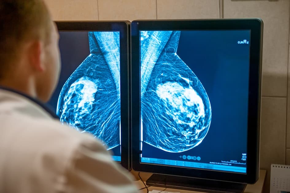
Artificial intelligence is transforming breast cancer screening, but new research reveals a significant performance gap when analyzing dense breast tissue. A study of 200,000 examinations from BreastScreen Norway found that AI-based cancer detection systems experience substantial performance degradation as breast density increases, highlighting a critical limitation in technology increasingly deployed across healthcare systems.
Study Findings: Performance Decline Across Density Categories
The research, published in European Radiology, analyzed screening examinations performed at eight Norwegian breast centers between 2010 and 2021 using Transpara version 2.1, a commercially available, CE-marked AI model from ScreenPoint Medical. The dataset included 1,235 screen-detected cancers and 368 interval cancers.
AI cancer detection performance declined substantially with increasing breast density. The area under the curve (AUC)—a measure of diagnostic accuracy—fell from 0.955 in the lowest-density breasts to 0.857 in the highest-density category when both screen-detected and interval cancers were considered together.
Interval cancer rates told an even more dramatic story. These cancers, diagnosed after a negative screening examination but before the next scheduled round, increased from 0.5 per 1,000 examinations in the lowest-density group to 3.1 per 1,000 in the highest-density group—a sixfold increase.
When researchers examined only screen-detected cancers, however, performance did not differ significantly between density groups. This suggests that interval cancers strongly influenced the overall performance decline, according to the study authors led by statistician Marthe Larsen of the Cancer Registry of Norway.
Equipment Variation and Measurement Inconsistency
The study revealed notable equipment-related variations in density classification. Among examinations performed using Siemens Healthineers equipment, 3.3% were placed in the highest volumetric density group, compared with 6.7% of examinations obtained using Hologic systems.
The researchers noted this difference does not prove the equipment caused the variation, as the systems were used at different centers and in different populations. Women screened using Hologic systems were more likely to live in urban areas, which may partly explain the variation.
Perhaps most striking, in 18.5% of examinations, the AI placed the left and right breasts in different density categories. This inconsistency raises practical questions about which measurement should determine a woman's screening pathway when implementing risk-stratified programs.
The Dense Breast Challenge
Dense breast tissue presents a dual challenge for cancer detection: it creates a masking effect that makes tumors harder to visualize on mammograms, and it represents an independent risk factor for developing breast cancer. The Norwegian findings demonstrate that AI systems designed to improve detection face similar limitations.
European breast screening is gradually moving away from uniform, age-based programs toward pathways that consider individual risk factors. In risk-stratified screening, a woman's risk factors influence how frequently she is screened and whether she receives mammography alone or supplemental imaging. The European Society of Breast Imaging (EUSOBI), for example, recommends MRI for women with extremely dense breasts.
Breast density is central to this transition. Automated density assessment could help determine who needs MRI, contrast-enhanced mammography (CEM), ultrasound, or more frequent screening. However, as the study demonstrates, the measurement must be reliable.
Work Remaining Before Clinical Implementation
The Norwegian findings identify substantial work still required before automated density assessment can guide personalized screening pathways. According to the researchers, European programs may need cross-vendor calibration, standardized thresholds, and a consistent method for converting breast-level measurements into a single clinical decision.
Major European trials have investigated supplemental screening for women with dense breasts, including the Dutch DENSE trial, which examined MRI, and the U.K. BRAID trial, which compared abbreviated MRI, CEM, and automated breast ultrasound.
The study's scope—200,000 examinations from a single national screening program using one AI system—provides robust evidence of the density-related performance variation, but also highlights the need for standardization work across different AI platforms, equipment manufacturers, and healthcare settings before automated density assessment can reliably guide screening decisions.
Sources
Companies in this article

Reporting & accountability
How we maintain accuracy
Health AI Central separates reported facts, analysis, and public-source data. Readers can review our standards, request a correction, or consult the relevant research methodology before relying on a claim.