
AI Reads Brain Scans to Predict Childhood Cancer Risk Without a Biopsy
A deep learning system achieved 82% accuracy classifying deadly pediatric brain tumors and their genetic signatures from routine MRI scans, potentially sparing young patients from invasive molecular testing.
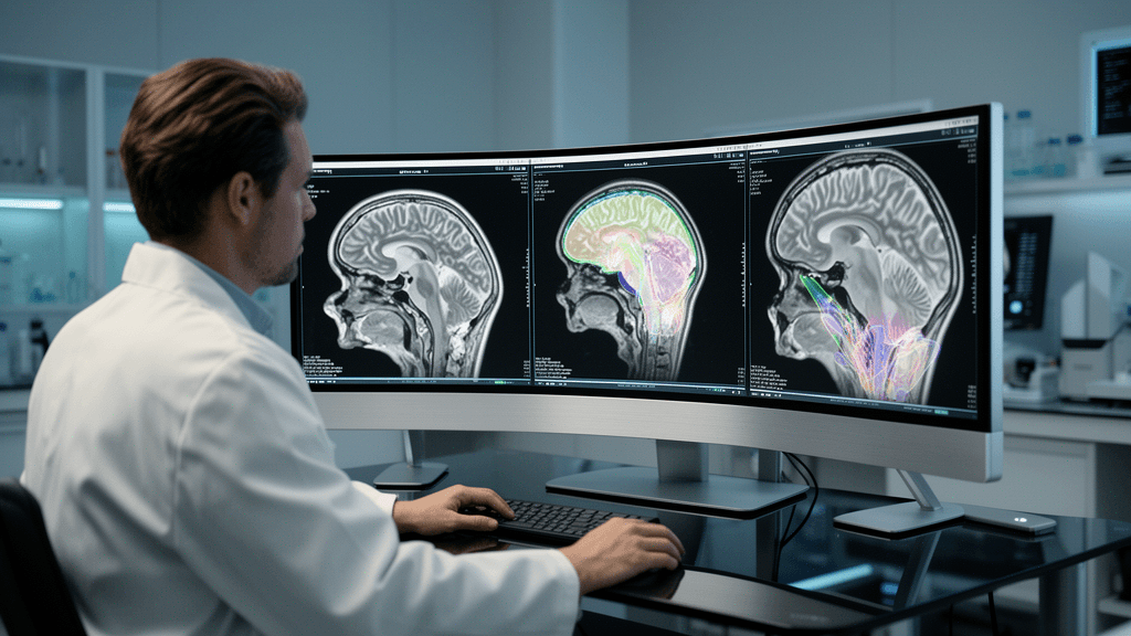
Pediatric brain cancer treatment may soon enter a new era where artificial intelligence determines tumor aggressiveness before surgery even begins. A multicenter study published in the Chinese Neurosurgical Journal reports that deep learning models analyzing preoperative MRI scans can accurately classify medulloblastoma molecular subgroups and predict high-risk genetic signatures with 77.50% median accuracy in external validation—potentially enabling noninvasive molecular risk stratification that currently requires invasive tissue sampling.
The research, encompassing 449 consecutive patients treated between January 2015 and June 2023 at two Beijing medical institutions, demonstrates how AI could transform treatment planning for the most common malignant pediatric brain tumor. When combined with clinical demographics and radiological features, the computational framework achieved 82.20% overall diagnostic accuracy, suggesting readiness for prospective clinical validation.
Dual-Stage Architecture Outperforms Earlier Approaches
The research team developed a two-stage system beginning with U-Net architecture for automated 3D tumor segmentation, achieving a mean Dice score of 0.91. After benchmarking four algorithms—VGG (65.03% precision), U-Net (72.00%), GoogLeNet (69.06%), and ResNet-50 (78.05%)—the investigators selected ResNet-50 as their classification core, designating it MB-CNN.
The 50-layer ResNet-50 architecture processed T1-weighted, T2-weighted, and contrast-enhanced T1-weighted MRI sequences from multiple scanner manufacturers. In the external validation cohort of 124 patients from Beijing Shijitan Hospital, MB-CNN achieved median accuracy of 77.50% (range 76.29% to 78.71%) classifying the four molecular subgroups: WNT, sonic hedgehog (SHH), Group 3, and Group 4.
This performance represents an advance over earlier medulloblastoma AI models, which achieved 70% to 85% accuracy but were constrained by limited cohort sizes, lack of external validation, and failure to predict high-risk genetic signatures.
Predicting Genetic Signatures That Determine Survival
The second stage targeted specific genetic alterations that drive treatment decisions. In 105 external validation patients with verified genetic signatures, the system demonstrated particularly strong performance predicting high-risk molecular features:
- MB-CNN_TP53 (sonic hedgehog tumors, n=44): AUC 0.93, accuracy 91.27%
- MB-CNN_MYC (Group 3 tumors, n=23): AUC 0.85, accuracy 83.56%
- MB-CNN_Chr11 (Group 4 tumors, n=38): AUC 0.88, accuracy 86.79%
These genetic signatures carry profound prognostic implications. According to the 2021 WHO classification framework, WNT tumors demonstrate >90% five-year overall survival, while Group 3 and Group 4 tumors with MYC amplification or chromosome 11 loss exhibit aggressive behavior and poor treatment response. SHH tumors show prognostic heterogeneity driven by TP53 mutation status.
Ground truth for model training derived from DNA methylation arrays, next-generation sequencing, whole-genome sequencing, and comparative genomic hybridization—the current gold standard requiring surgical tissue samples.
Multimodal Integration Boosts Accuracy
The hybrid model integrated MB-CNN outputs with patient demographics and conventional radiological features assessed by two neuroradiologists with high inter-reader agreement (kappa 0.842-0.864, P<0.001). This multimodal approach increased overall diagnostic accuracy from 77.50% to 82.20%, suggesting that combining AI-derived imaging biomarkers with clinical parameters yields superior performance compared to imaging-only approaches.
The study population included 249 males and 200 females with mean age 13.55 years, spanning from infants under 3 years (n=14, mean 1.86 years) through adults aged 18-49 years (n=68, mean 28.67 years). The training cohort comprised 325 patients from Beijing Tiantan Hospital.
Clinical Implications for Precision Pediatric Oncology
This dual-stage framework could enable preoperative molecular risk stratification that guides therapeutic intensity decisions without diagnostic delays associated with tissue-based molecular profiling. Accurate classification before surgery may help clinicians avoid over-treatment toxicities in low-risk patients while ensuring adequate therapeutic intensity for high-risk genetic signatures.
The investigators note that conventional radiological evaluation introduces subjective assessment and misclassification risk, whereas the automated deep learning approach provides standardized, reproducible molecular predictions from routine imaging studies performed on 1.5T and 3.0T systems from multiple manufacturers including Philips Healthcare, Siemens Healthineers, GE Healthcare, and Toshiba Medical Systems.
The research demonstrates potential clinical viability of AI systems addressing unmet needs in molecular subtyping for specialized pediatric neuro-oncology applications. The accuracy gains achieved through multimodal integration suggest that hybrid clinical-imaging models may offer advantages over imaging-only approaches.
Sources
Companies in this article

Follow Health AI Central
Keep up with health AI where you already read
On LinkedIn
The day’s strongest health AI stories in your feed, one or two each weekday.
On Google
Choose Health AI Central as a preferred source, and Google is more likely to show our reporting in Top Stories.
Reporting & accountability
How we maintain accuracy
Health AI Central separates reported facts, analysis, and public-source data. Readers can review our standards, request a correction, or consult the relevant research methodology before relying on a claim.